National Neuroimaging Facility
The National Neuroimaging Facility, sponsored by the Department of Biotechnology, Govt. of India, came into existence in the year of 2006. The main purpose of this National Facility is to facilitate/support cutting edge brain imaging research. The facility is equipped with three state-of-the-art equipments including:
- 3T Magnetic Resonance Imaging (MRI)Scanner
- Electroencephalography (EEG)
- Evoked Response Potential Recording (ERP)
Magnetic Resonance Imaging (MRI):
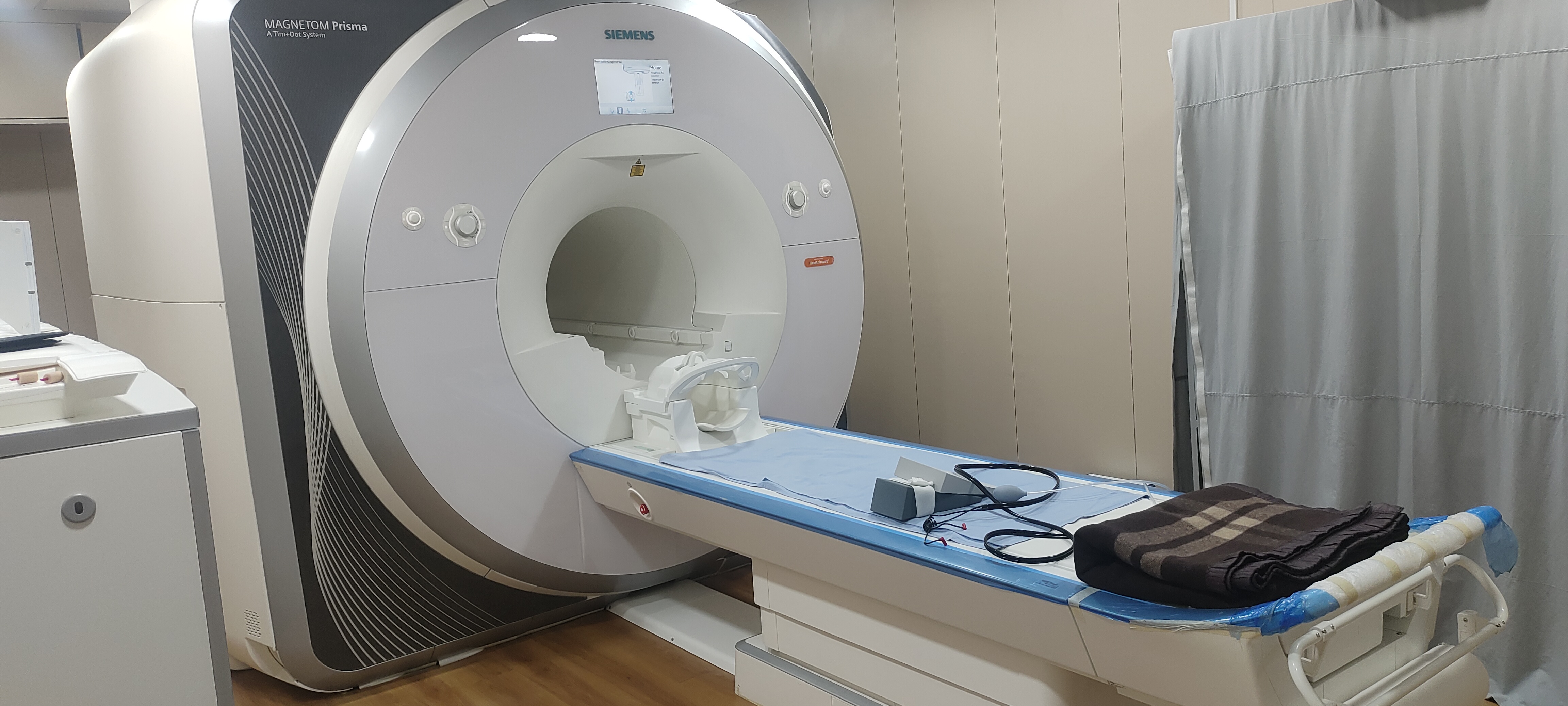
NBRC is equipped with Siemens 3 Tesla Magnetom Prisma MRI Scanner (3T MRI) with stronger magnet having 80/200 gradient performance with latest parallel transmit technology and highest coil element density. Being state of the art Scanner, it can produce complex images by maintaining consistency & reproducibility of research at the same time. It ensures that results are far more reproducible and of excellent in quality due to its Tim 4G technology, reducing the time of scanning by 30 to 40 percent as compared to conventional MRIs. It contributes to identifying the brain connectivity, body physiology, organ morphometry, tissue metabolism, and quantitative MRI.
Following are its key modalities that significantly contributes to research:-
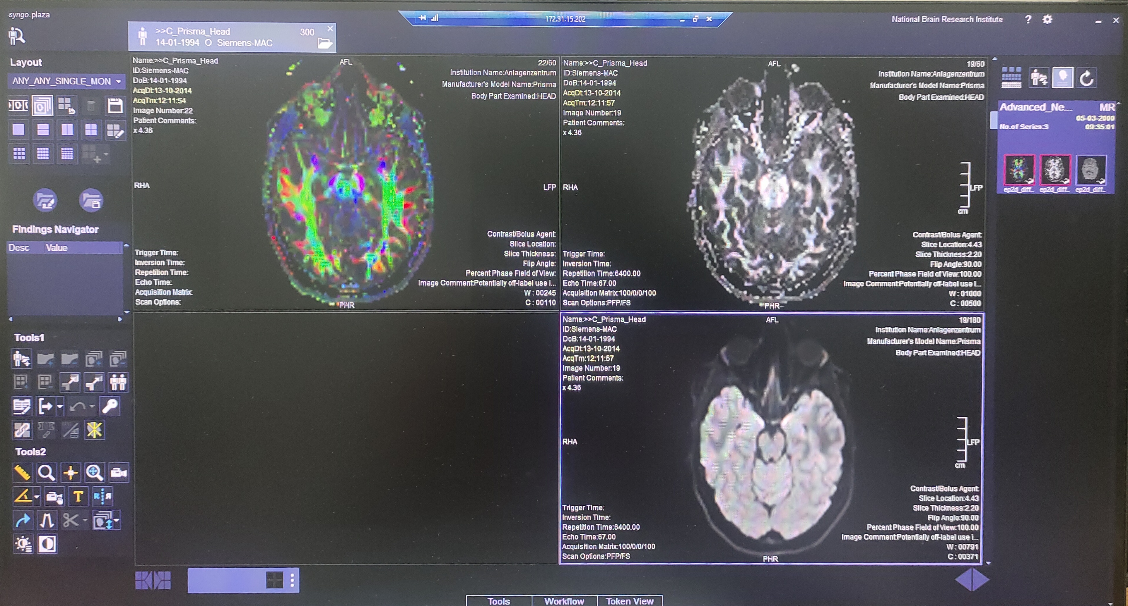
- Whole Brain DTI Tractography (Brain Connectivity)
- C-Spine DTI Tractography
- Body Physiology (Cardiac Flow Imaging)
- Organ Morphometry (High resolution knee with excellent FatSat)
Advanced applications of DWI, SWI, resting state fMRI, simultaneous 128 Channel EEG, dynamic angiography, perfusion, and DSI acquisition contributes considerably in research. Magnetom Prisma allows us to visualize details in the finest anatomical structures and reveals valuable functional information. Besides, we can develop our own applications and sequences - adapted to our research needs. The following Coils are part of the Magnetom Prisma facility:-
- Head/Neck 64 Channel Coil
- Head/Neck 32 Channel Coil
- Spine 32 Channel Coil
- Flex 4 Channel Shoulder/Elbow and joints Coil
- Body 18 Channel Coil
- Knee Coil 15 Channel Coil
- 31P/1H Head MRS Coil
- 19F Head Volume Coil
- 23 Na1H Sodium Head Coil
- 1H Phased Array Coil for Monkey Head
The facility is being used for performing structural, metabolic (multinuclear, e.g. proton and phosphorous) and functional MRI (fMRI). In addition to understanding brain function and clinical research, the National Neuroimaging Facility at NBRC closely interacts with leading imaging centers within the country and across the globe.
Contact:
Mr. Jitender Ahlawat, Technical Officer - B, MRI Facility
jitender[at]nbrc[dot]ac[dot]in
0124-2845318 (318)
Electroencephalography (EEG) is a test that measures and records the electrical activity of the brain. Special sensors are attached to the head and connected by wires to a computer. The computer records brain’s electrical activity on the screen or on paper as wavy lines. Certain conditions, such as epilepsy, dementia, consciousness and narcolepsy (sleeping disorder) can be studied by EEG.
Evoked Response Potential Recording (ERP) is an electrical potential recorded from the nervous system of a human or other animal following presentation of a stimulus. Evoked potential amplitudes tend to be low, ranging from less than a microvolt to several microvolts
The Neuroimaging and Neurospectroscopy laboratory at NBRC is working on the metabolic analysis of different neurodegenerative disorders (e.g. Alzheimer, Parkinson etc) using the MRS technique. The clinical research using this technique is aimed at identifying biomarkers, quantitative stress measurements from age-matched normal subjects and patients. This is accomplished by applying novel MRS pulse sequences and signal processing schema on scans of human subjects. Figure 1 shows a gradual decrease in the glutathione molecule, an important anti-oxidant in the brain, with aging and in different clinical conditions. This is the first report of the quantitative measurement of oxidative stress using a non-invasive state-of-the-art technique
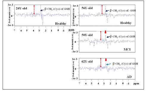
Figure 1 GSH levels in the right frontal cortex in various cases: (A) healthy female (26Y old); (B) healthy female (56Y old); (C) MCI female (50Y old) and (D) Probable AD female patient (62Y old) using MEGA-PRESS pulse sequence in a 3T MRI scanner. The decrease in GSH content is indicated by red arrow.
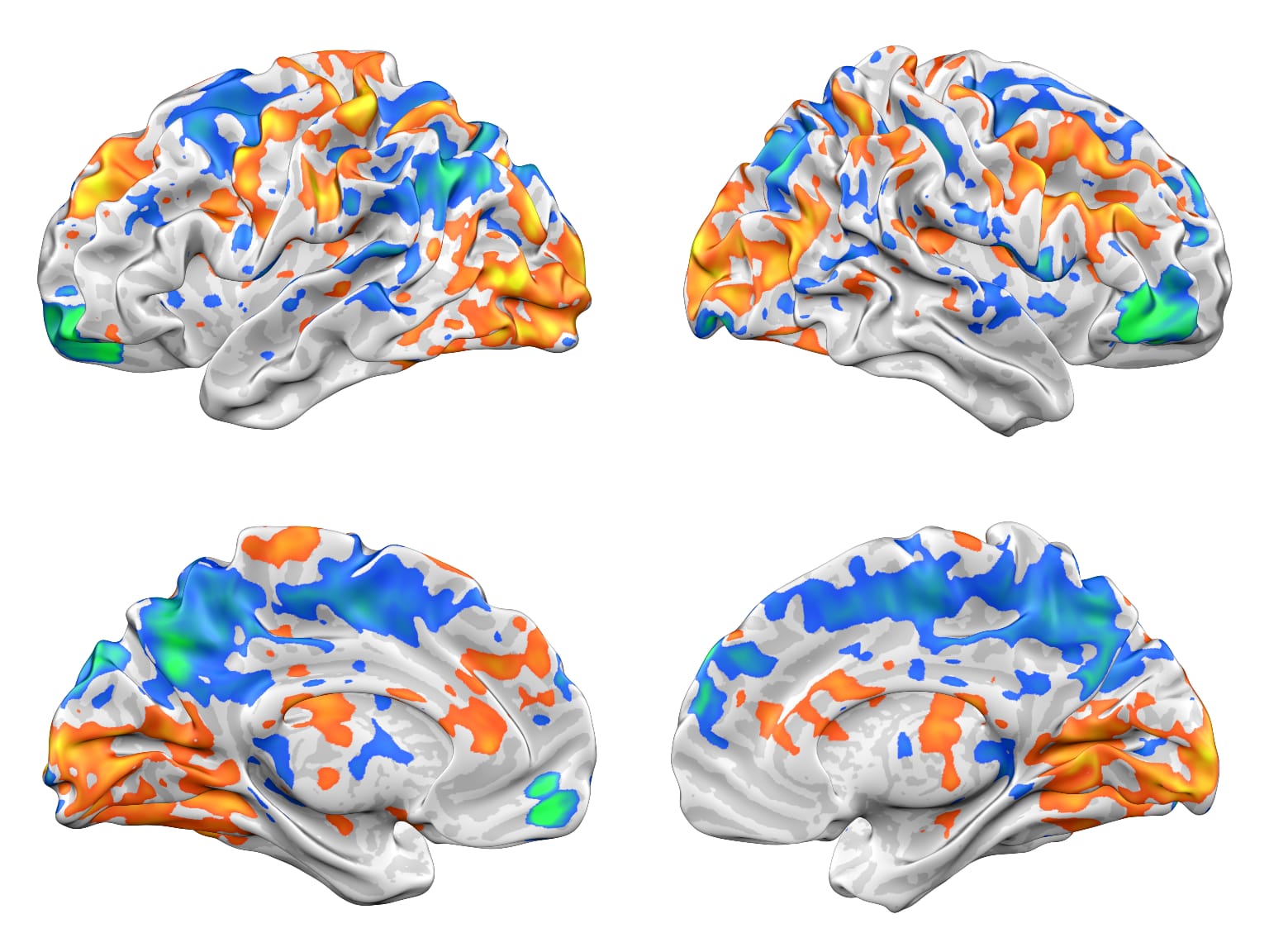
The Speech and Language Laboratory (SALLY) Research in the Speech and Language. SALLY is focused on unraveling the cortical pathways involved in Hindi-English biscriptal adults and children (Figure 2).
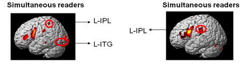
Figure 2 shows how skilled readers use different cortical networks.
The Computational Neuroscience and Neuroimaging Laboratory works on diagnostic and therapeutic applications with a translational medicine aspect. The unit works on imaging-based diagnosis of neurodegenerative disease and planning pulsed radiotherapy and chemotherapy for treating brain tumours. In collaboration with clinical centres and medical institutes, the lab also pursues the delineation of flow dynamics of blood, CSF and progenitor cells in brain, as well as localization of electrogenic foci in refractory epilepsy using EEG, ERP, fMRI, MRS and DTI (diffusion tensor imaging) (Figure 3).
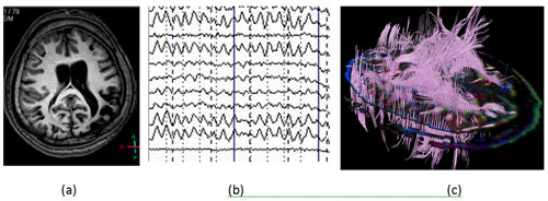
Figure 3 (a) Structural MRI scan of brain with dysplasia in an epileptic patient. (b) EEG recording of the brain which enables one to obtain more accurate localization of the electrical foci (c) Diffusion tensor image (DTI) of the brain of epileptic patient from which the conductivity tensor image (CTI) of the brain is obtained, the latter image enables accurate localization of the electrogenic epileptic focus in brain tissue, based on the conductivity image obtained earlier.
